43 monocular microscope diagram with label
Label the microscope — Science Learning Hub In this interactive, you can label the different parts of a microscope. Use this with the Microscope parts activity to help students identify and label the main parts of a microscope and then describe their functions. Drag and drop the text labels onto the microscope diagram. Diagrams of binocular microscope with labels? - Answers A monocular microscope has only one eyepiece while a binocular microscope has two eyepieces with different lenses. Binocular microscopes are more popular today than the monocular...
Microscopy: Intro to microscopes & how they work (article) - Khan Academy This picture isn't a plain light micrograph; it's a fluorescent image of a specially prepared plant where various parts of the cell were labeled with tags to make them glow. However, this kind of cellular complexity and beauty is all around us, whether we can see it or not.

Monocular microscope diagram with label
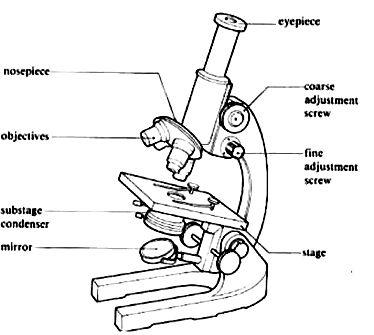
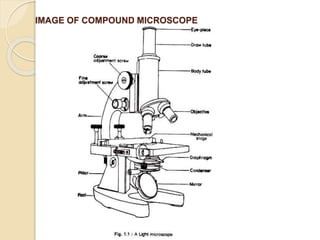
Solved ASSIGNMENT 1) Draw a neat diagram of a monocular - Chegg Question: ASSIGNMENT 1) Draw a neat diagram of a monocular microscope and label the different parts. Trace diagrammatically the path of the light rays from the mirror to the eye through the microscope 2) Draw neatly the various equipment and glassware used in a microbiology laboratory. 3) Explain Scanning & Transmission Electron Microscope. Labeling the Parts of the Microscope | Microscope World Resources Microscope World explains the parts of the microscope, including a printable worksheet for schools and home. Need Asssistance? 800-942-0528. Microscope Blog ... Labeling the Parts of the Microscope. This activity has been designed for use in homes and schools. Each microscope layout (both blank and the version with answers) are available as PDF ... Parts of a microscope with functions and labeled diagram - Microbe Notes Figure: Diagram of parts of a microscope There are three structural parts of the microscope i.e. head, base, and arm. Head - This is also known as the body. It carries the optical parts in the upper part of the microscope. Base - It acts as microscopes support. It also carries microscopic illuminators.
Monocular microscope diagram with label. Parts of Stereo Microscope (Dissecting microscope) - labeled diagram ... Stereo microscopes (also called Dissecting microscope) are branched out from other light microscopes for the application of viewing "3D" objects. These include substantial specimens, such as insects, feathers, leaves, rocks, sand grains, gems, coins, and stamps, etc. Functionally, a stereo microscope is like a powerful magnifying glass. Diagram and labels of a monocular microscope? - Answers Diagram of monocular microscope with a neat labbling? The photo below may help you with this: What is the difference between a monocular microscope and a binocular microscope? A... Microscope Parts, Types & Diagram | What is a Microscope? There are many illustrations available for the diagram of a light microscope. The essential parts include the head, base, arms, lenses, and lights. In diagrams, one would see the head always... Compound Microscope Parts - Labeled Diagram and their Functions Labeled diagram of a compound microscope Major structural parts of a compound microscope There are three major structural parts of a compound microscope. The head includes the upper part of the microscope, which houses the most critical optical components, and the eyepiece tube of the microscope.
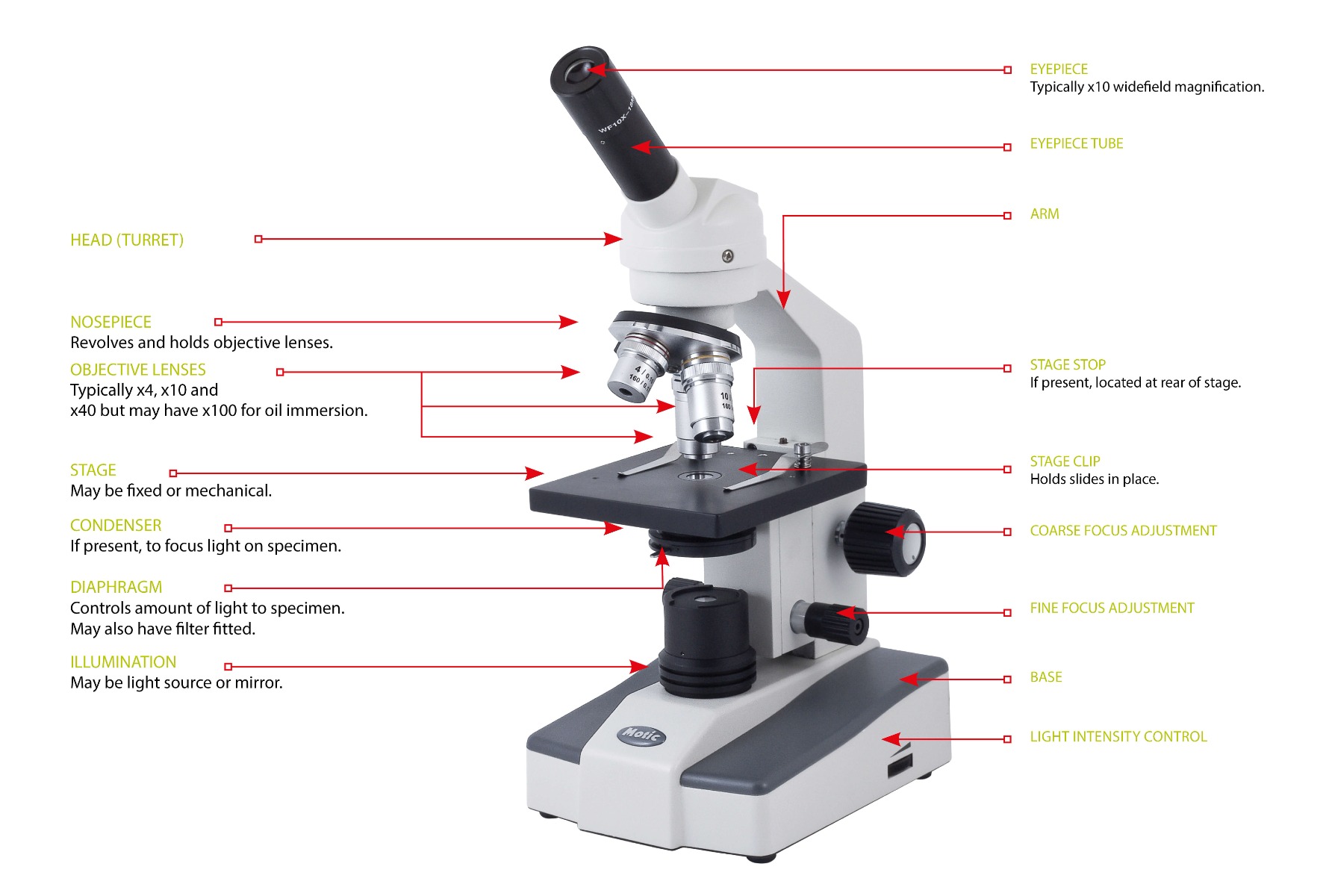
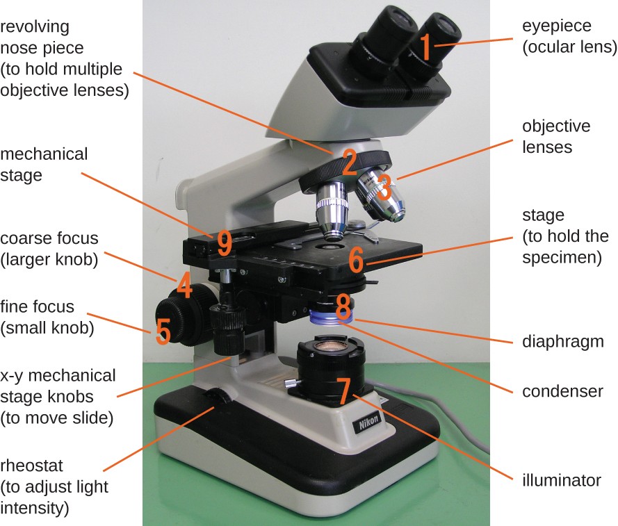
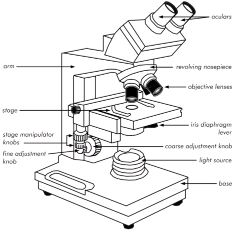
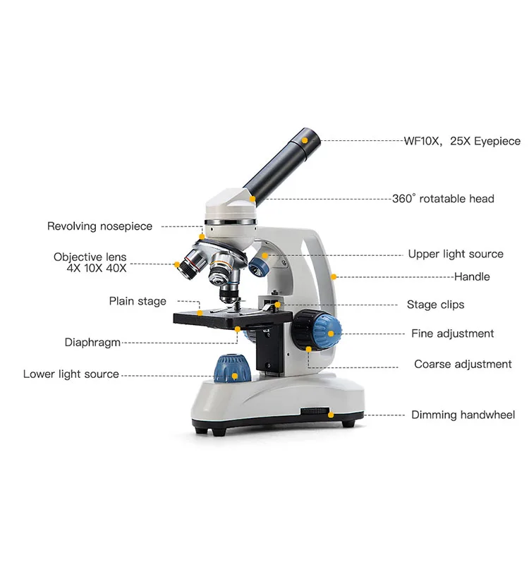
Compound microscope - their parts and function - Microscopy4kids 2. Eyepiece (10x) and Objective lenses (4x, 10x, 40x, 100x) are two major optical parts of a microscope. 3. Total magnification power is calculated by multiplying the magnification of the eyepiece and objective lens. 4. A proper immersion oil helps oil lens achieve an ideal magnification or resolution. 5. Microscope - The compound microscope | Britannica The compound microscope. The limitations on resolution (and therefore magnifying power) imposed by the constraints of a simple microscope can be overcome by the use of a compound microscope, in which the image is relayed by two lens arrays. One of them, the objective, has a short focal length and is placed close to the object being examined.It is used to form a real image in the front focal ... How to Label a Binocular Microscope | Sciencing Label the arm. Identify the stage. The stage is located beneath the objective lenses and holds the specimen at the appropriate distance from the objective lens. The stage is equipped with stage clips to hold the specimen in place. Label the stage. Locate the iris diaphragm underneath the stage. Monocular - Wikipedia Monocular sizes. As with binoculars and telescopes, monoculars are primarily defined by two parameters: magnification and objective lens diameter, for example, 8×30 where 8 is the magnification and 30 is the objective lens diameter in mm (this is the lens furthest from the eye).An 8× magnification makes the distant object appear to be 8 times larger at the eye.
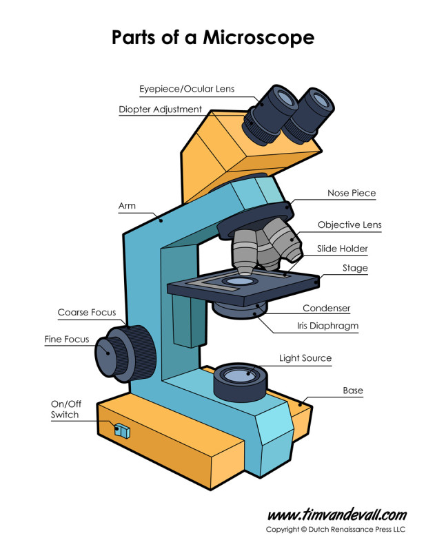
PDF Basic Microscopy Laboratory Exercises - CDC 1. Correctly identify various parts of a brightfield microscope. Exercises: 1. Label the correct parts of a brightfield microscope on the graphic on the following page. 2. Identify the following parts of a brightfield microscope on the bench microscope you are using: A. Objectives B. Condenser (Iris) diaphragm C. Coarse adjustment 16 Parts of a Compound Microscope: Diagrams and Video Once you have an understanding of the parts of the microscope it will be much easier to navigate around and begin observing your specimen, which is the fun part! The 16 core parts of a compound microscope are: Head (Body) Arm. Base. Eyepiece. Eyepiece tube. monocular labeling Diagram | Quizlet monocular labeling STUDY Learn Flashcards Write Spell Test PLAY Match Gravity + − Created by jt8sdmx Key concepts: Light Stage Clips Objective Lenses Terms in this set (8) eyepiece the lens at the top that you look through. it enlarges the image usually by 10X. body tube Connects the eyepiece to the objective lenses nosepiece Binocular Microscope Anatomy - Parts and Functions with a Labeled Diagram Now, I will discuss the details anatomy of the light compound microscope with the labeled diagram. Why it is called binocular: because it has two ocular lenses or an eyepiece on the head that attaches to the objective lens, this ocular lens magnifies the image produced by the objective lens. Binocular microscope parts and functions
Compound Microscope Parts, Function, & Diagram | What is a ...
Solved (1)Draw a neat diagram | Chegg.com This problem has been solved! You'll get a detailed solution from a subject matter expert that helps you learn core concepts. See Answer. Question: (1)Draw a neat diagram of a monocular microscope and label the different parts. Trace diagrammatically the path of the light rays from the mirror to the eye through the.

267 Eyepiece Science Stock Vector Illustration and Royalty ...
Compound Microscope Parts, Functions, and Labeled Diagram Compound Microscope Definitions for Labels Eyepiece (ocular lens) with or without Pointer: The part that is looked through at the top of the compound microscope. Eyepieces typically have a magnification between 5x & 30x. Monocular or Binocular Head: Structural support that holds & connects the eyepieces to the objective lenses.

Parts of a Microscope with Their Functions • Microbe Online
Microscope Parts and Functions It also allows the specimen to be labeled, transported, and stored without damage. Stage: The flat platform where the slide is placed. Stage clips: Metal clips that hold the slide in place. Stage height adjustment (Stage Control): These knobs move the stage left and right or up and down.

Compound microscope - their parts and function - Microscopy4kids
Parts of the Microscope with Labeling (also Free Printouts) A microscope is one of the invaluable tools in the laboratory setting. It is used to observe things that cannot be seen by the naked eye. Table of Contents 1. Eyepiece 2. Body tube/Head 3. Turret/Nose piece 4. Objective lenses 5. Knobs (fine and coarse) 6. Stage and stage clips 7. Aperture 9. Condenser 10. Condenser focus knob 11. Iris diaphragm
Using Your Microscope: Buying a Microscope – Koi & Aquarium ...
16 Essential Microscope Parts: Names, Functions & Labeled Diagram Microscope Parts Labeled Diagram The principle of the Microscope gives you an exact reason to use it. It works on the 3 principles. Magnification Resolving Power Numerical Aperture. Parts of Microscope Head Base Arm Eyepiece Lens Eyepiece Tube Objective Lenses Nose Piece Adjustment Knobs Stage Aperture Microscopic Illuminator Condenser Lens

AmScope M149C-PS50-WM Compound Monocular Microscope, WF10x ...
Parts of a microscope with functions and labeled diagram - Microbe Notes Figure: Diagram of parts of a microscope There are three structural parts of the microscope i.e. head, base, and arm. Head - This is also known as the body. It carries the optical parts in the upper part of the microscope. Base - It acts as microscopes support. It also carries microscopic illuminators.
Microscope Diagram Labeled, Unlabeled and Blank | Parts of a ...
Labeling the Parts of the Microscope | Microscope World Resources Microscope World explains the parts of the microscope, including a printable worksheet for schools and home. Need Asssistance? 800-942-0528. Microscope Blog ... Labeling the Parts of the Microscope. This activity has been designed for use in homes and schools. Each microscope layout (both blank and the version with answers) are available as PDF ...

Guide to understand microscope parts, names, functions & diagram
Solved ASSIGNMENT 1) Draw a neat diagram of a monocular - Chegg Question: ASSIGNMENT 1) Draw a neat diagram of a monocular microscope and label the different parts. Trace diagrammatically the path of the light rays from the mirror to the eye through the microscope 2) Draw neatly the various equipment and glassware used in a microbiology laboratory. 3) Explain Scanning & Transmission Electron Microscope.
Monocular microscope hi-res stock photography and images - Alamy

2000X Mikroskop Biologis dengan Kamera Digital USB 2.0MP ...

Compound Microscope – Diagram (Parts labelled), Principle and ...

Simple Microscope- Definition, Principle, Magnification ...

A Compound Monocular Optical Microscope | Download Scientific ...

Parts Of A Microscope And Their Function

Swift Compound Monocular Microscope Kit for Kids Students ...

AmScope M148C-E Compound Monocular Microscope Instruction ...
Parts of a Microscope - Annotated Diagram

Compound Microscope Principle, Parts, Diagram Definition ...
Instruments of Microscopy | Microbiology | | Course Hero
Glossary of terms used in microscopy – Quekett Microscopical Club

Cost-effective Monocular Biological Microscope AS1

Dissecting Stereo Microscope Parts and Functions

HOw to draw light or compound microscope step by step / Microscope diagram
What are the parts of the brightfield microscope? PreLab 3.8

Basic Malaria Microscopy (part I and II) (WHO - OMS, 1991, 72 ...
Swift-sw150 Rotatable Professional Microscope 1000x Monocular Microscope For Kids Biological Microscope - Buy Biological Microscope,Light ...

40X to 800X Multi-View Monocular Compound Microscope + 0.3MP ...

GEMKO LABWELL Nose Single Lens Compound Baby Microscope, LED ...

Microscopy forum - MicrobeHunter Microscopy Magazine ...

Compound Microscope Parts, Functions, and Labeled Diagram ...

Amazon.com: BestScope BS-2000A Basic Monocular Compound ...
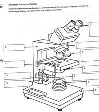
Answered: Microscope Structure and Function… | bartleby
Introduction to microscopy

compound monocular light microscope Diagram | Quizlet
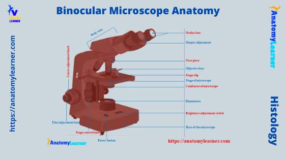
Binocular Microscope Anatomy - Parts and Functions with a ...

Parts of a microscope with functions and labeled diagram

Chapter 5 Cells Units Of Life - Lessons - Blendspace

Microscope Diagram Labeled, Unlabeled and Blank | Parts of a ...

Label the microscope — Science Learning Hub

Types of Microscopes: Definition, Working Principle, Diagram ...

Labeling the Parts of the Microscope | Microscope World Resources

monocular labeling Diagram | Quizlet

Compound Microscope Parts, Functions, and Labeled Diagram ...
Post a Comment for "43 monocular microscope diagram with label"